Impacted third molars: Winter and Pell & Gregory
What you need to know
- Third molars, if they come in, appear roughly between the ages of 17 and 21. Some stay inside the bone: they're said to be retained or impacted.
- Winter classifies them by how the third molar is tilted compared with the second: vertical (parallel), mesioangular (tilted forward), horizontal (lying down) and distoangular (tilted backward). There's also the inverted type (with the crown facing down).
- In studies, the most common are mesioangular and vertical; which one comes out on top depends on the population. In a study of 1,055 molars, mesioangular made up 41.8%.
- Pell and Gregory look at 2 things. The space in front of the mandibular ramus: Class I (plenty of space), Class II (a bit short of space) and Class III (no space).
- And the depth compared with the second molar: position A (level with its chewing surface), B (between that surface and the cervical line of the tooth) and C (below the cervical line).
- Problems they can cause: pericoronitis (infection of the gum covering the tooth), caries on the back surface of the second molar, pain, swelling and wearing away of neighboring roots (resorption).
Winter's classification
| Type | How the tooth sits |
|---|---|
| Vertical | Parallel to the second molar |
| Mesioangular | Tilted forward, toward the second molar |
| Horizontal | Lying down, perpendicular to the second molar |
| Distoangular | Tilted backward, toward the ramus |
| Inverted | The crown faces down |
Terms in plain words
Impacted toothA tooth that doesn't fully erupt because something blocks it (also called a retained tooth).
PericoronitisInflammation or infection of the gum covering a molar that's erupting.
Mandibular ramusThe vertical part of the mandible, behind the molars.
MesioangularTilted forward (mesially).
For the exam
A lower third molar is tilted forward, resting against the second molar. What is it called according to Winter?
Show answerHide answer
Mesioangular.
Sources
- Santos KK, et al. Prevalence of Mandibular Third Molars According to the Pell & Gregory and Winter Classifications. J Maxillofac Oral Surg. 2022;21(2):627–633. View source
- American Dental Association (MouthHealthy): eruption charts. View source
More from Oral Surgery & Implants
Extraction
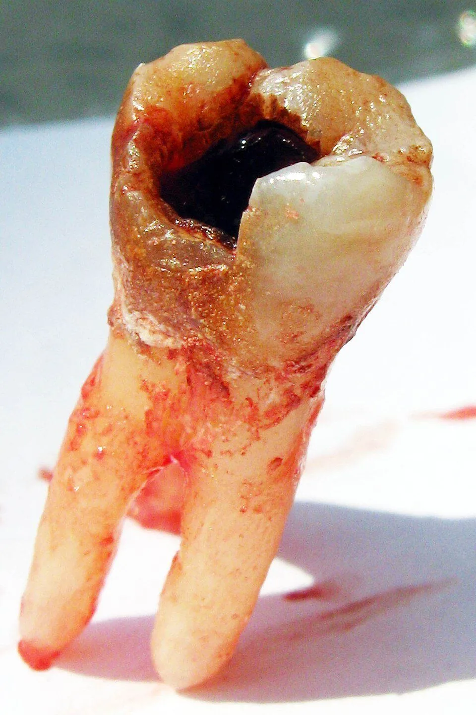
Simple extraction, step by step
In a simple extraction, you break the periodontal ligament and expand the socket with elevators, and then remove the tooth with forceps.
Healing
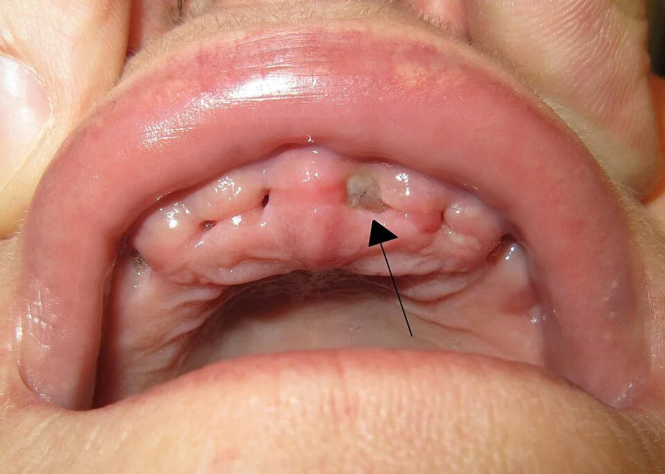
Healing after an extraction
After an extraction, the socket fills with blood clot → granulation tissue → immature bone → mature bone.
Implants
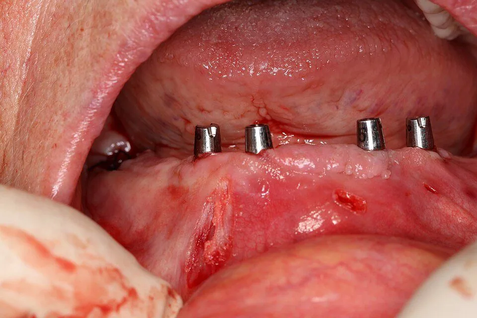
Osseointegration
Osseointegration is the direct, structural and functional connection between living bone and the surface of an implant that bears load (the force of chewing).