Zones of the dental pulp
What you need to know
- Odontoblast layer: odontoblasts in a row, right against the predentin. Their processes extend into the dentinal tubules.
- Cell-free zone (zone of Weil): almost no cells; it has thin nerve fibers (without myelin, the layer that insulates other nerves), fibroblast processes and capillaries (the thinnest vessels).
- Cell-rich zone: fibroblasts and progenitor cells (pulp stem cells) that can replace odontoblasts.
- Pulp core: the center, with the largest blood vessels and nerves.
- The fibroblast is the most abundant cell in the pulp.
Terms in plain words
OdontoblastCell that makes dentin; it lives at the edge of the pulp.
PredentinNewly formed dentin that has not mineralized yet.
FibroblastCell that forms the fibers of connective tissue; the most abundant cell in the pulp.
For the exam
Which zone lies just beneath the odontoblast layer?
Show answerHide answer
The cell-free zone, or zone of Weil.
Sources
- Grawish ME. Human dental pulp stem/stromal cells in clinical practice. World J Stem Cells. 2024;16(2):54–57. View source
- Farges JC, et al. Dental Pulp Defence and Repair Mechanisms in Dental Caries. Mediators Inflamm. 2015:230251. View source
More from Histology & Embryology
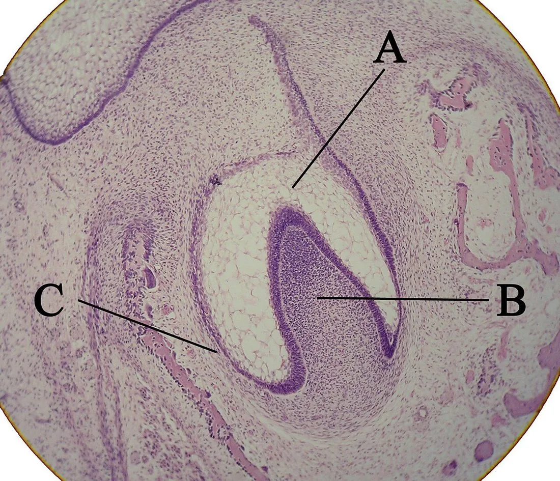
Odontogenesis
Odontogenesis: how a tooth forms
From the 6th week of intrauterine life, the tooth germ goes through 3 stages: bud, cap and bell.
Dentin
Primary, secondary and tertiary dentin
There are 3 types of dentin depending on when and why it forms: primary, secondary and tertiary.
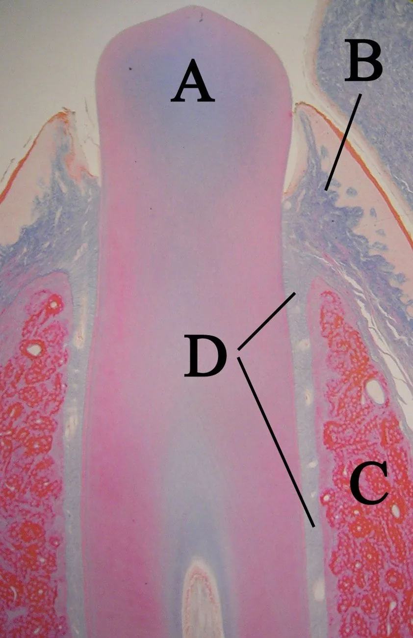
Root
Root formation: Hertwig's epithelial root sheath
The root forms thanks to Hertwig's epithelial root sheath: a double layer of epithelium that grows apically (toward where the tip of the root will end up) and defines the shape of the root.