Odontogenesis: how a tooth forms
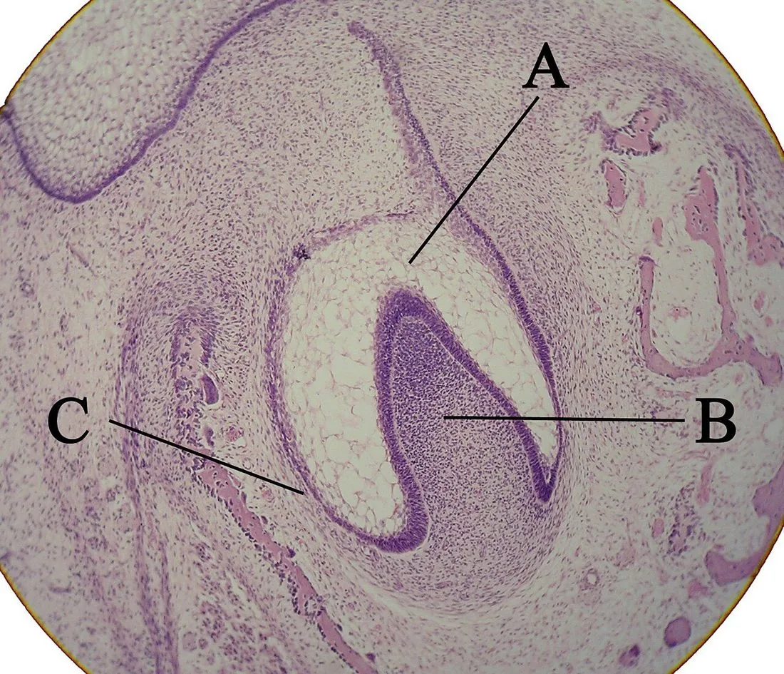
A · Enamel organB · Dental papillaC · Dental follicle
Real histological section of a tooth germ seen under the microscope.Photo: Wikimedia Commons user · CC BY-SA 3.0, Wikimedia CommonsWhat you need to know
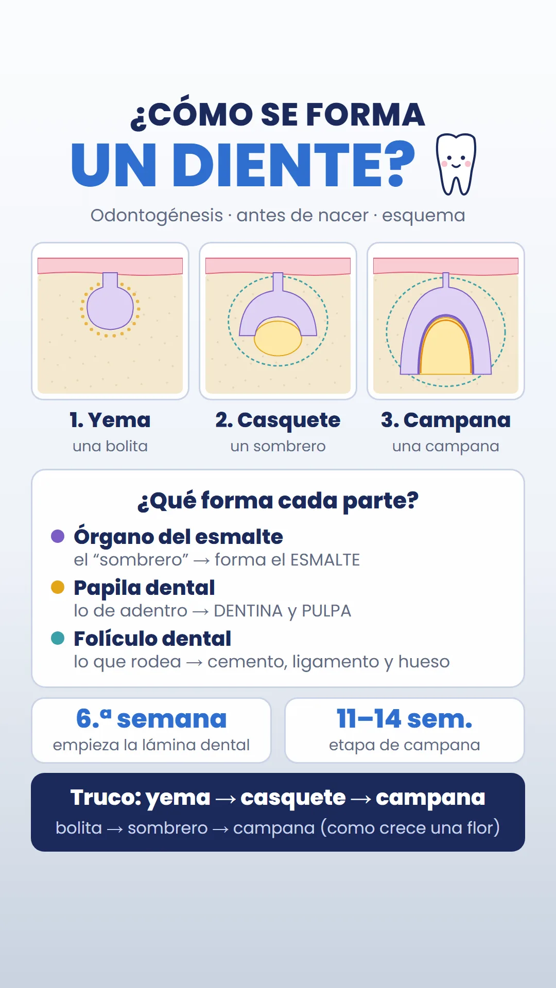
- Initiation: around the 6th week, the epithelium of the mouth forms the dental lamina.
- Bud: the lamina forms a little ball surrounded by condensed mesenchyme (a tightly packed group of cells that will later form the papilla and the follicle).
- Cap: the bud hollows out like a hat over the dental papilla.
- Bell: the shape of the crown is defined, and the ameloblasts and odontoblasts appear.
- Then come mineralization, root formation (Hertwig's epithelial root sheath) and eruption.
- The exact weeks vary a little from book to book. A 2025 review places the cap stage between weeks 8 and 10 and the bell stage between weeks 11 and 14.
Terms in plain words
Enamel organThe epithelial “hat”. Its ameloblasts make the enamel.
Dental papillaWhat lies inside the hat. It forms the dentin (odontoblasts) and the pulp.
Dental follicleWhat surrounds everything. It forms the cementum, the periodontal ligament and the alveolar bone.
For the exam
Which structure is dentin derived from?
Show answerHide answer
From the dental papilla, through the odontoblasts. Tip: bud → cap → bell = little ball → hat → bell.
Sources
- Novacescu D, et al. The Morphogenesis, Pathogenesis, and Molecular Regulation of Human Tooth Development: A Histological Review. Int J Mol Sci. 2025;26(13):6209. View source
- Nanci A. Ten Cate's Oral Histology. Elsevier (textbook).
More from Histology & Embryology
Pulp
Zones of the dental pulp
From the outside in, the pulp has 4 zones: odontoblast layer, cell-free zone (of Weil), cell-rich zone and pulp core.
Dentin
Primary, secondary and tertiary dentin
There are 3 types of dentin depending on when and why it forms: primary, secondary and tertiary.
Root
Root formation: Hertwig's epithelial root sheath
The root forms thanks to Hertwig's epithelial root sheath: a double layer of epithelium that grows apically (toward where the tip of the root will end up) and defines the shape of the root.