Root formation: Hertwig's epithelial root sheath
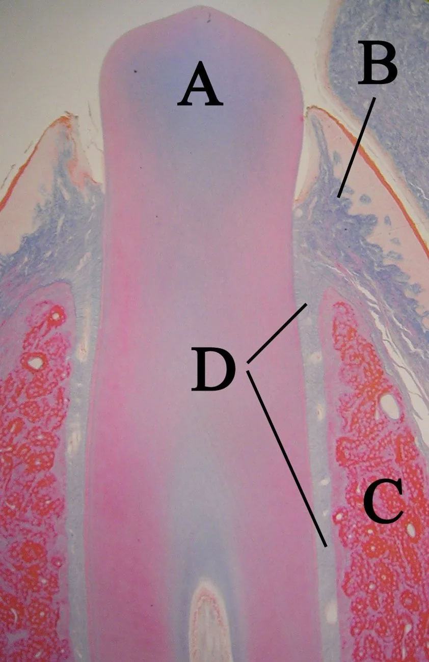
A · ToothB · GingivaC · BoneD · Periodontal ligament
Real histological section of a tooth with its root inside the bone.Photo: Dozenist · CC BY-SA 3.0, Wikimedia CommonsWhat you need to know
- It appears once the crown has formed. It arises from the cervical loop: the junction of the inner and outer epithelium of the enamel organ.
- The sheath causes the cells of the papilla to become odontoblasts, which form the dentin of the root.
- When that first dentin mineralizes, the sheath breaks up. Cells from the follicle come into contact with the dentin and become cementoblasts, which form the cementum.
- What is left of the sheath stays in the periodontal ligament as the epithelial rests of Malassez.
Terms in plain words
Cervical loopThe rim of the enamel organ where its inner and outer layers meet.
CementoblastCell that forms the cementum covering the root.
Epithelial rests of MalassezClusters of epithelial cells that remain in the periodontal ligament.
For the exam
What remains in the periodontal ligament when Hertwig's root sheath breaks up?
Show answerHide answer
The epithelial rests of Malassez.
Sources
- Fate of HERS during Tooth Root Development (research article). View source
- Evolution and Development of Hertwig's Epithelial Root Sheath (review). View source
More from Histology & Embryology
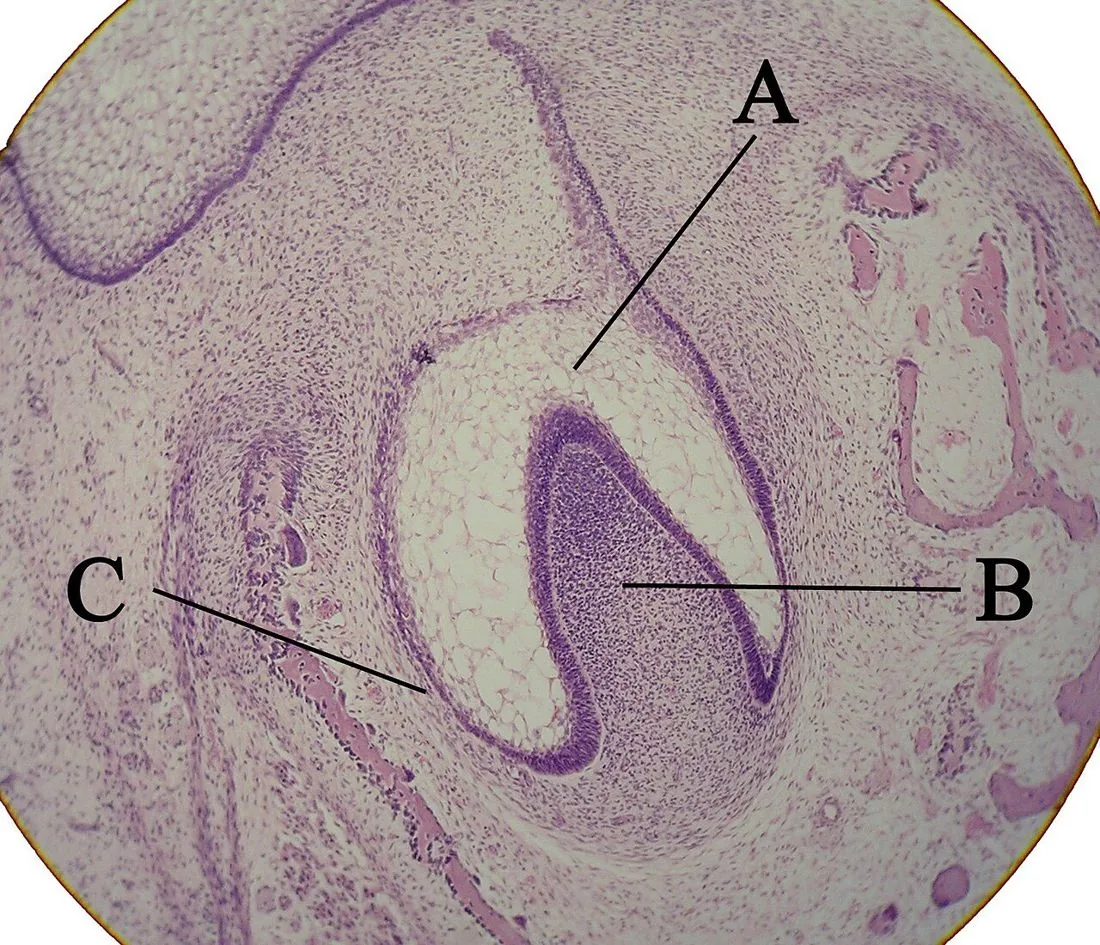
Odontogenesis
Odontogenesis: how a tooth forms
From the 6th week of intrauterine life, the tooth germ goes through 3 stages: bud, cap and bell.
Pulp
Zones of the dental pulp
From the outside in, the pulp has 4 zones: odontoblast layer, cell-free zone (of Weil), cell-rich zone and pulp core.
Dentin
Primary, secondary and tertiary dentin
There are 3 types of dentin depending on when and why it forms: primary, secondary and tertiary.