The trigeminal nerve and its 3 divisions
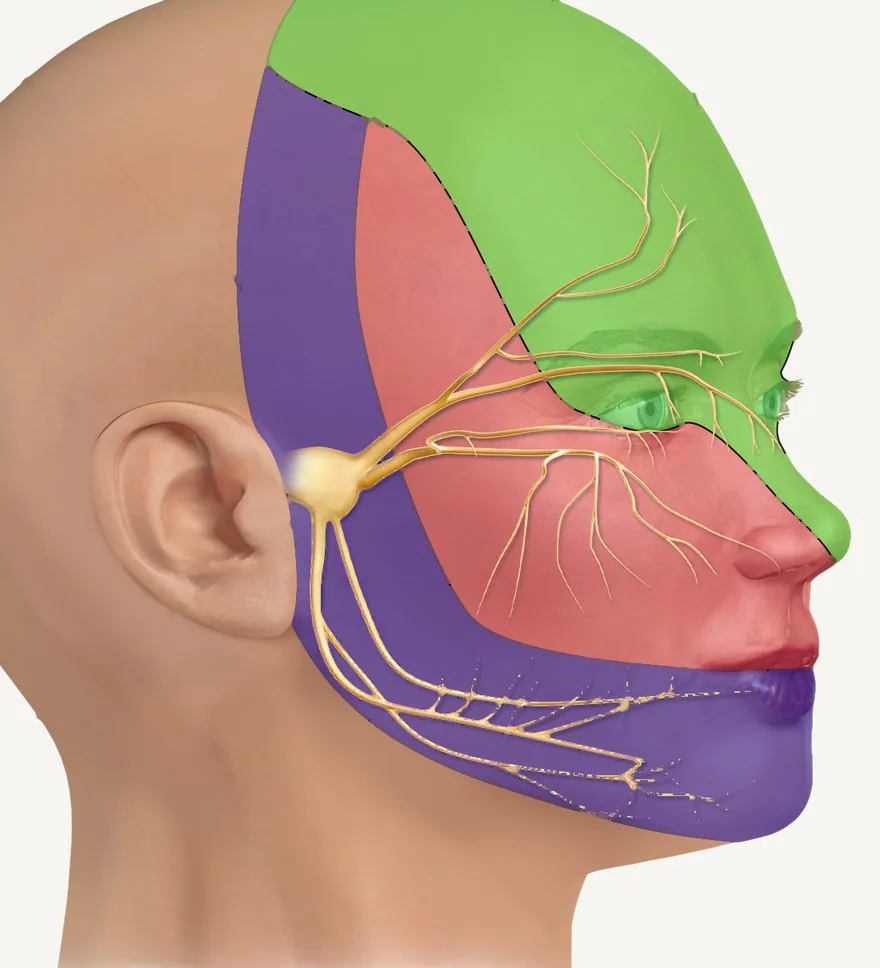
V1 · ophthalmicV2 · maxillaryV3 · mandibular
The 3 trigeminal zones: green is the ophthalmic (V1), red the maxillary (V2) and purple the mandibular (V3). The angle of the mandible is left uncolored because it isn't trigeminal territory.Photo: BruceBlaus / Blausen Medical (modified: labels removed) · CC BY-SA 4.0, Wikimedia CommonsWhat you need to know
- It's a mixed nerve: almost all of it is sensory (touch, pain and temperature from the face, teeth and mouth), and it has a motor part that runs only in the mandibular division.
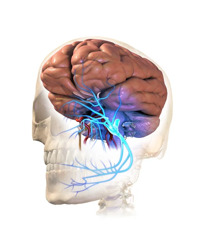
- The sensory fibers have their cell bodies in the trigeminal ganglion (Gasserian ganglion), in the middle cranial fossa, in a depression of the temporal bone called the trigeminal cave. The 3 divisions come out from there.
- V1 · Ophthalmic: forehead, front of the scalp, upper eyelid, cornea and dorsum of the nose. It leaves the skull through the superior orbital fissure.
- V2 · Maxillary: lower eyelid, cheek, ala of the nose, upper lip, upper teeth and gingiva, and palate. It exits through the foramen rotundum.
- V3 · Mandibular: lower lip, chin, the skin in front of the ear and the temple, lower teeth and gingiva, and touch from the front 2/3 of the tongue. It exits through the foramen ovale.
- V3 is the only motor division: it moves the 4 muscles of mastication (masseter, temporalis, medial and lateral pterygoid), and also the mylohyoid, the anterior belly of the digastric, the tensor veli palatini and the tensor tympani.
- The angle of the mandible is not trigeminal territory: that skin is innervated by the cervical nerves C2 and C3. It's a classic exam question.
- In dentistry: the superior alveolar nerves (from V2) give sensation to the upper teeth and the inferior alveolar nerve (from V3) to the lower ones. That's why, for the lower teeth, you block the inferior alveolar nerve.
- Trigeminal neuralgia: brief attacks of very intense, electric-shock-like pain, almost always on one side only, in the territory of one or more divisions. They can be triggered by something as simple as touching your face or brushing your teeth.
The 3 divisions at a glance
| Division | Exits through | Area |
|---|---|---|
| V1 · Ophthalmic (sensory) | Superior orbital fissure | Forehead, upper eyelid, cornea, dorsum of the nose |
| V2 · Maxillary (sensory) | Foramen rotundum | Cheek, lower eyelid, upper lip, upper teeth, palate |
| V3 · Mandibular (mixed: also motor) | Foramen ovale | Lower lip, chin, lower teeth, tongue (touch) |
Terms in plain words
Cranial nerveA nerve that comes directly out of the brain. There are 12 pairs; the trigeminal is CN V.
Mixed nerveOne that carries sensation and also moves muscles.
Gasserian ganglionThe trigeminal ganglion: where the cell bodies of its sensory neurons are.
NeuralgiaPain that starts in a nerve and follows its path.
For the exam
What is the only division of the trigeminal nerve with motor fibers, and through which foramen does it leave the skull?
Show answerHide answer
The mandibular division (V3). It exits through the foramen ovale and moves the muscles of mastication.
Sources
- TeachMeAnatomy. The Trigeminal Nerve (CN V): course, divisions. View source
- Kenhub. Trigeminal nerve (CN V): anatomy, function and branches. View source
- Truini A, et al. Trigeminal neuralgia. J Headache Pain. 2015 (review). View source
More from Dental Anatomy
FDI
FDI notation: how teeth are numbered
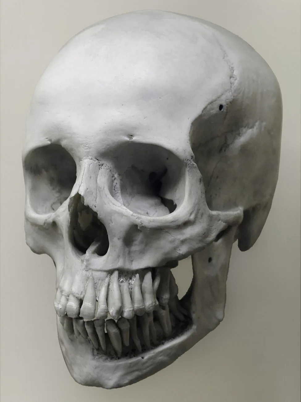
Each tooth is named with 2 digits: the first tells you the quadrant and the second, the position counting from the midline.
Eruption
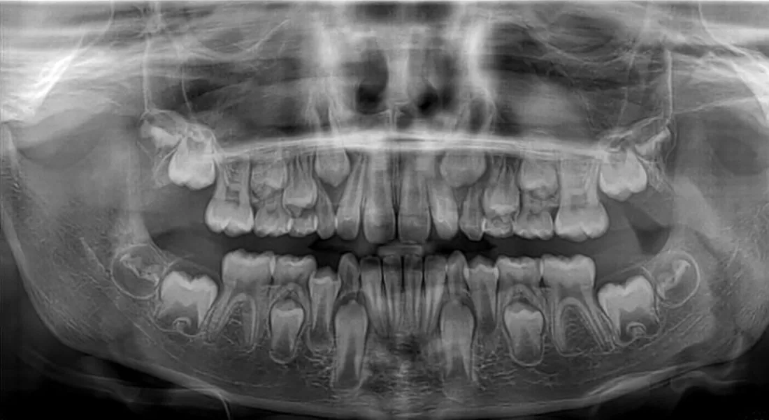
Eruption chronology: when each tooth comes in
The first baby tooth comes in at around 6 months; at age 3 there are 20 primary teeth, and by around age 21 all 32 permanent teeth have come in.
Successors
Successional and non-successional teeth
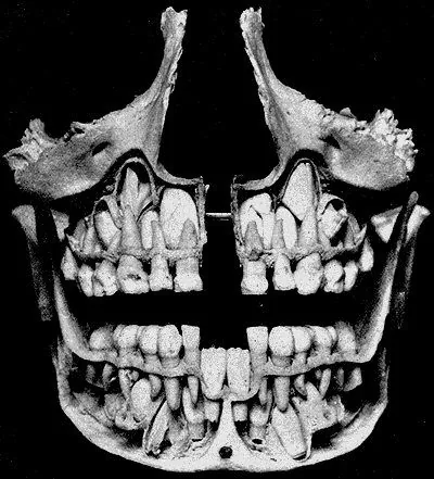
The permanent molars don't replace any baby tooth: that's why they're called non-successional, and there are 12 of them.