Odonto Fácil: dental school made easy, with real photos
Dentistry notes with official sources, terms in plain words and games to practice FDI notation and what they'll ask you on the exam.
Tap a subject to see its notes. Use the arrows or swipe to see them all.
Each note has a real photo or an exact diagram, what you need to know, the terms in plain words, an exam question and its sources.
37 notes
FDI notation: how teeth are numbered
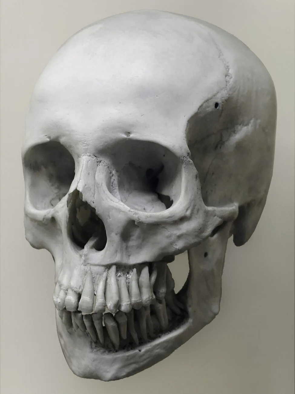
Each tooth is named with 2 digits: the first tells you the quadrant and the second, the position counting from the midline.
Eruption chronology: when each tooth comes in
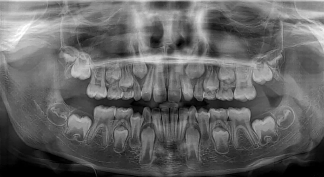
The first baby tooth comes in at around 6 months; at age 3 there are 20 primary teeth, and by around age 21 all 32 permanent teeth have come in.
Successional and non-successional teeth
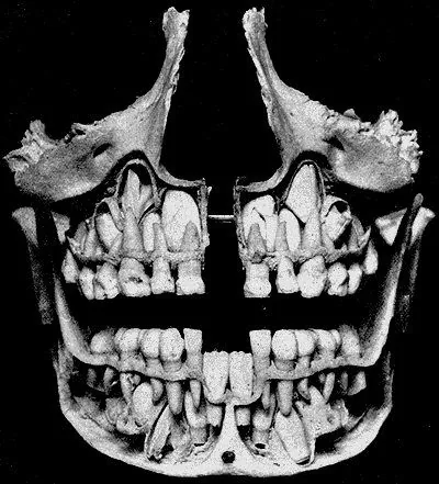
The permanent molars don't replace any baby tooth: that's why they're called non-successional, and there are 12 of them.
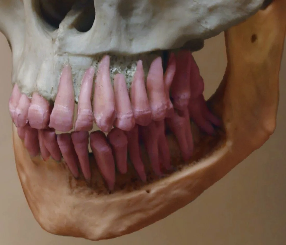
Upper or lower? How to identify a tooth
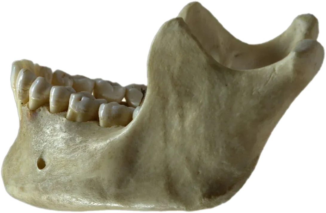
To tell whether a tooth is upper or lower, look at the roots and the size of the crown: upper molars usually have 3 roots and lower ones, 2.
Upper first molar and the cusp of Carabelli
The upper first molar has 4 main cusps and, often, a small extra one on the palatal surface (the one facing the palate): the cusp of Carabelli.
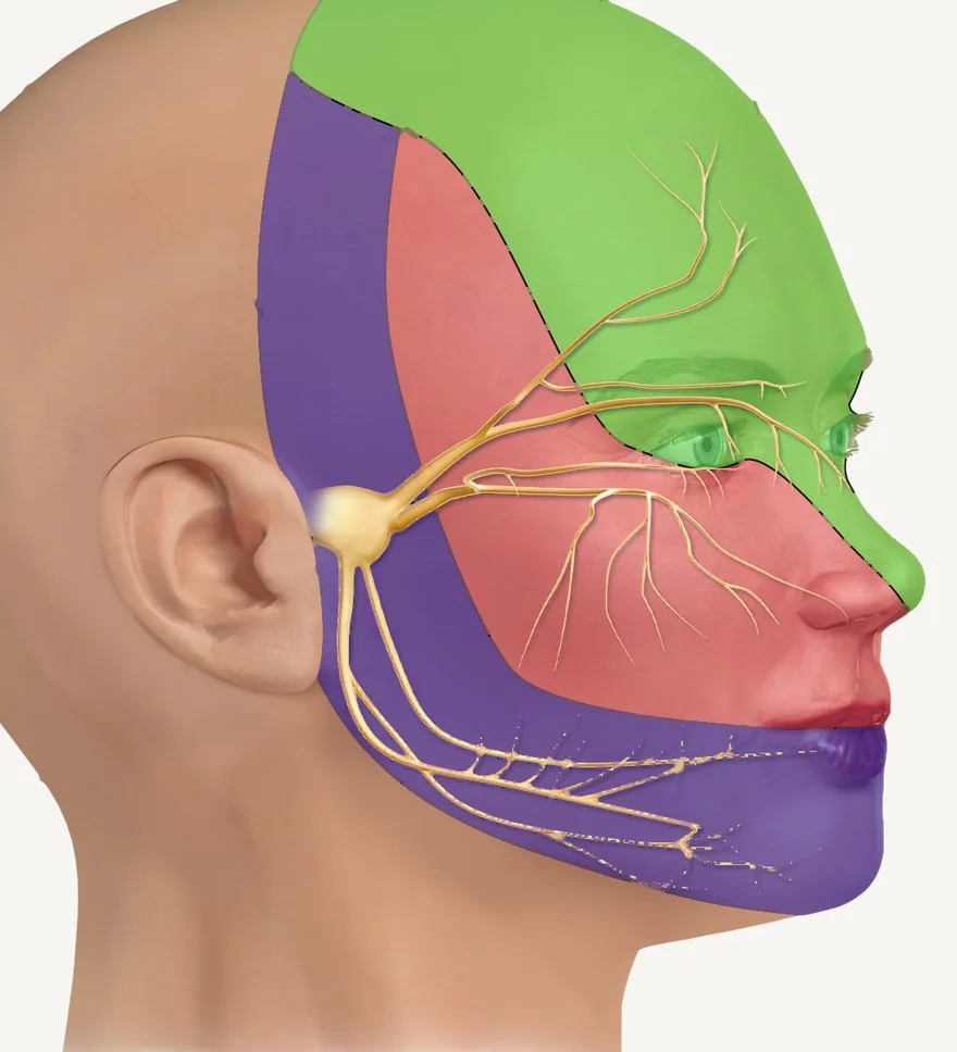
The trigeminal nerve and its 3 divisions
The trigeminal is the fifth cranial nerve: it gives sensation to the whole face and splits into 3 divisions, ophthalmic (V1), maxillary (V2) and mandibular (V3).
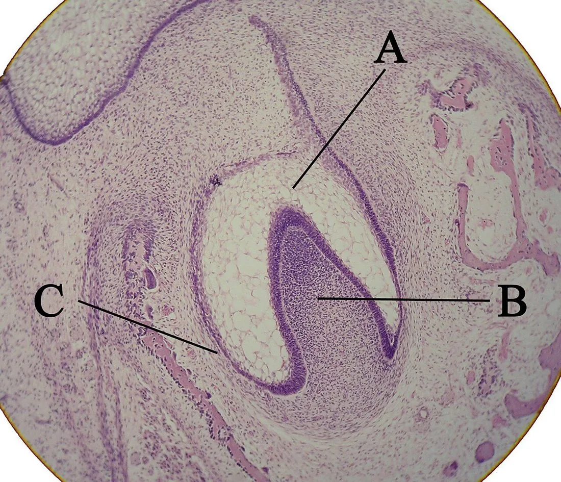
Odontogenesis: how a tooth forms
From the 6th week of intrauterine life, the tooth germ goes through 3 stages: bud, cap and bell.
Zones of the dental pulp
From the outside in, the pulp has 4 zones: odontoblast layer, cell-free zone (of Weil), cell-rich zone and pulp core.
Primary, secondary and tertiary dentin
There are 3 types of dentin depending on when and why it forms: primary, secondary and tertiary.
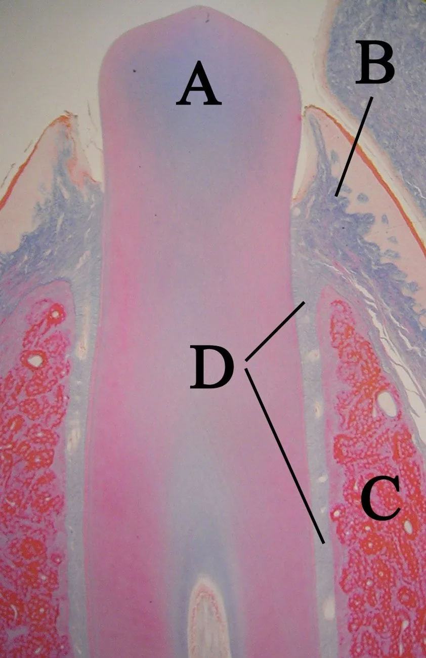
Root formation: Hertwig's epithelial root sheath
The root forms thanks to Hertwig's epithelial root sheath: a double layer of epithelium that grows apically (toward where the tip of the root will end up) and defines the shape of the root.
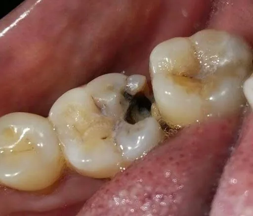
Stages of caries
Caries progresses from the outside in: enamel → dentin → pulp. While it is only in the enamel, it can still be remineralized.
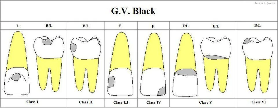
Black's classification
G. V. Black classified caries and cavities by where they are on the tooth: Classes I to V, and Class VI was added later.
Factors behind caries
For caries to occur, 4 factors have to come together: a susceptible tooth, bacteria, sugars and time.
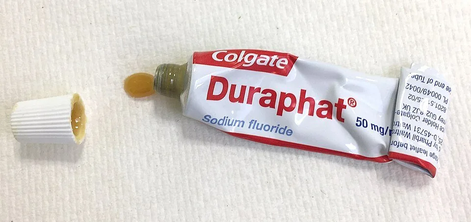
How fluoride protects
Fluoride helps with remineralization, makes enamel more resistant to acid and can reduce the acid made by bacteria.
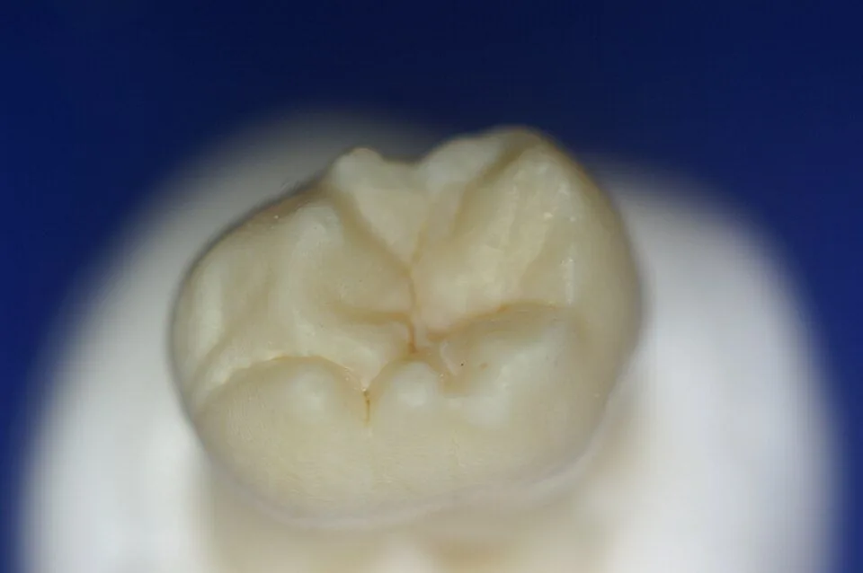
Pit and fissure sealants
A sealant is a protective coating painted onto the pits and fissures of the back teeth so bacteria do not get trapped.
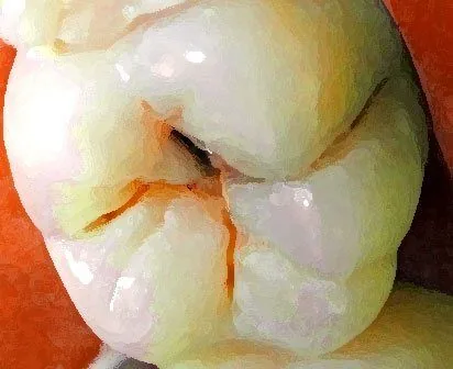
Composite resin restoration, step by step
Composite resin sticks to the tooth by adhesion: the tooth is etched, a bonding agent is applied and the resin is placed in thin layers that are hardened with light.
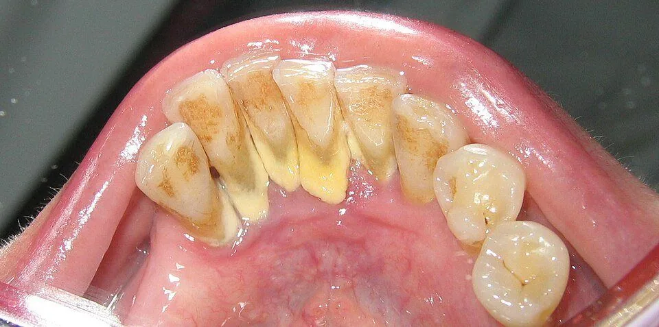
How calculus forms
Calculus is bacterial plaque that has mineralized with the calcium and phosphate in saliva.
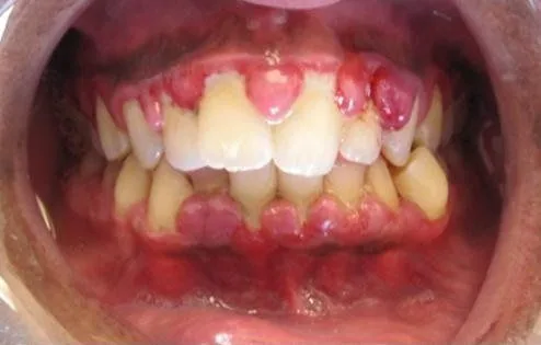
Gingivitis vs. periodontitis
Gingivitis inflames only the gingiva. Periodontitis also destroys the ligament and the bone that support the tooth.
Tooth mobility grades
Mobility is measured in grades according to how much the crown moves: from 0 (normal) to 3 (severe).
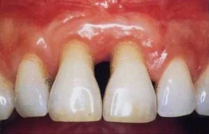
Gingival recession: Miller and Cairo classifications
Recession is when the gingival margin drops below the cementoenamel junction (the line where the enamel ends) and leaves the root exposed.
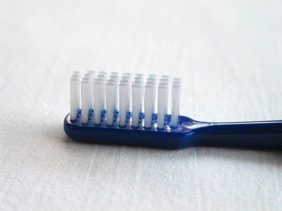
The Bass brushing technique
In the Bass technique, the brush goes at 45° toward the gums, with the bristles pointing at the sulcus, and you make short, gentle movements.
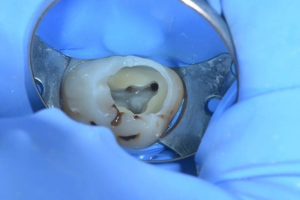
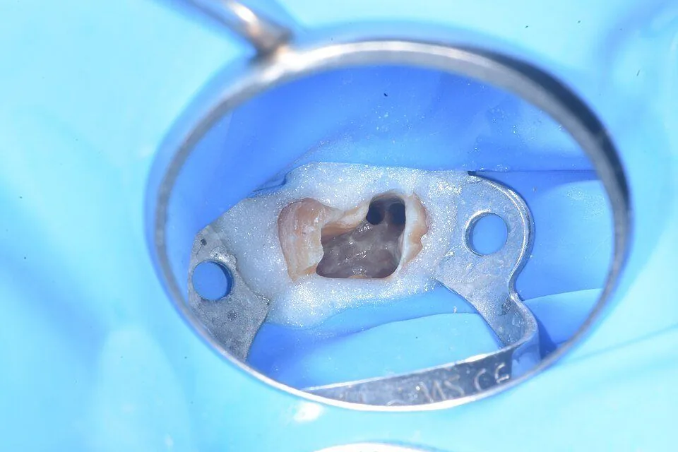
Root canal treatment, step by step
In root canal treatment, the inflamed or infected pulp is removed, the inside is cleaned and disinfected, and it is filled and sealed.
Pulp diagnosis: tests and terminology (AAE)
To find out the condition of the pulp, you compare the tooth with its neighbors using cold, percussion, palpation and radiographs.
Reversible vs. irreversible pulpitis
In reversible pulpitis, the pain goes away when the stimulus is removed. In irreversible pulpitis, the pain is spontaneous or lingers after the stimulus.
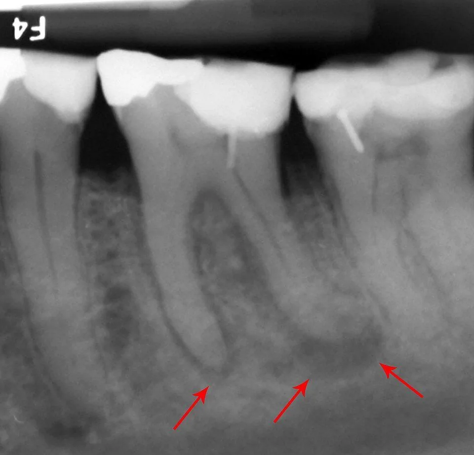
How an abscess forms
A periapical abscess develops when the pulp dies and the infection exits through the tip of the root into the bone.
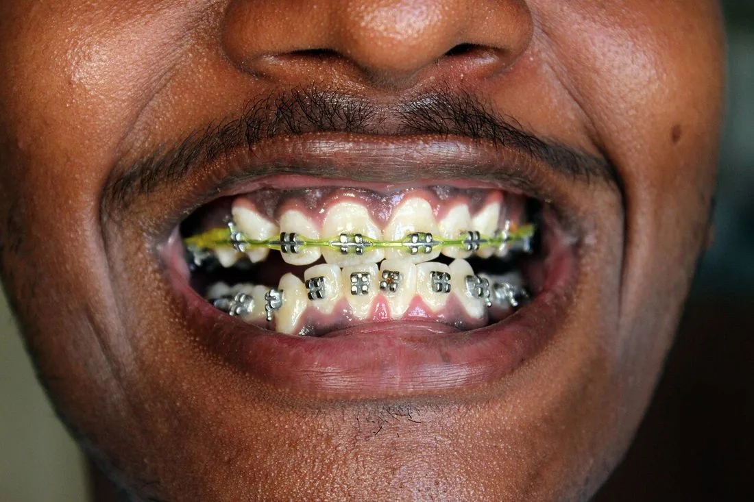
Tooth movement in orthodontics (pressure and tension)
The tooth doesn't force its way through the bone: the bone rebuilds itself around it. It's resorbed on one side and formed on the other.
Angle's classification
Angle classifies the bite by how the mesiobuccal cusp of the upper first molar fits with the buccal groove of the lower first molar.
Overjet, overbite and types of bite
Besides the Angle class, you look at how the front teeth meet: the overjet (forward) and the overbite (downward).
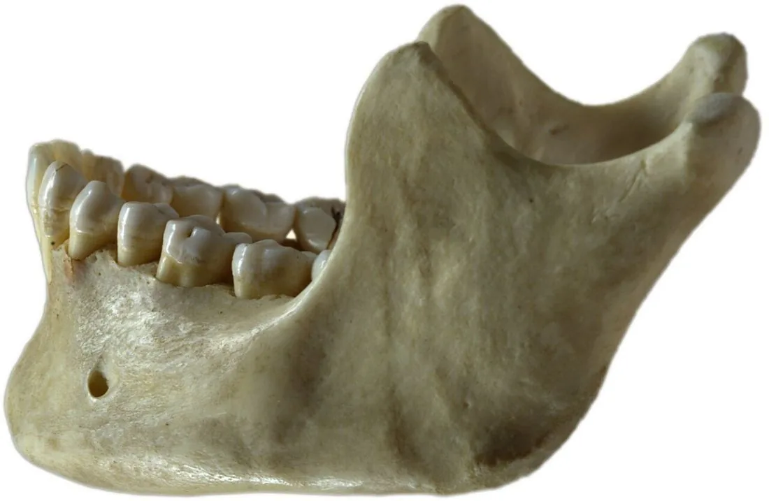
Canine guidance and group function
In canine guidance, when you move your lower jaw to one side, only the canines touch and the back teeth separate.
How local anesthesia works
Local anesthesia switches off the nerve signal for a while: the pain doesn't reach the brain, but the person stays awake.
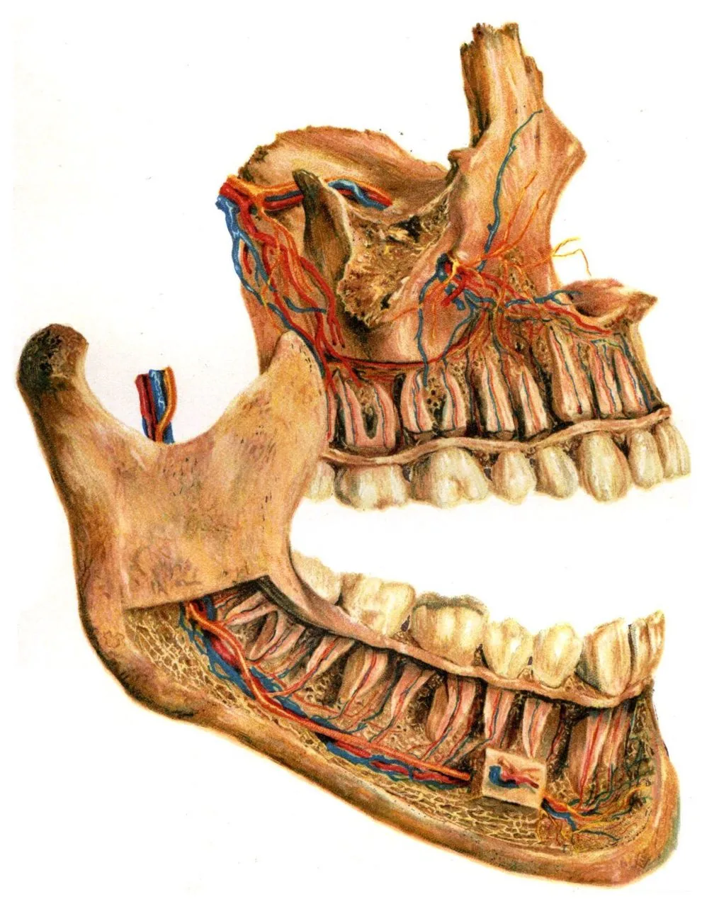
Inferior alveolar nerve block
It numbs the lower teeth on that side, the lip and chin, and almost always the tongue too.
Which nerve do you numb for each tooth?
To numb a tooth, you need to know which nerve gives it sensation: for the upper teeth it's the superior alveolar nerves, and for the lower teeth, the inferior alveolar nerve.
Safe anesthesia: aspiration, dosage and complications
Before injecting, you aspirate so you don't put the anesthetic into a blood vessel, you inject slowly, and you never go over the maximum dose based on weight.
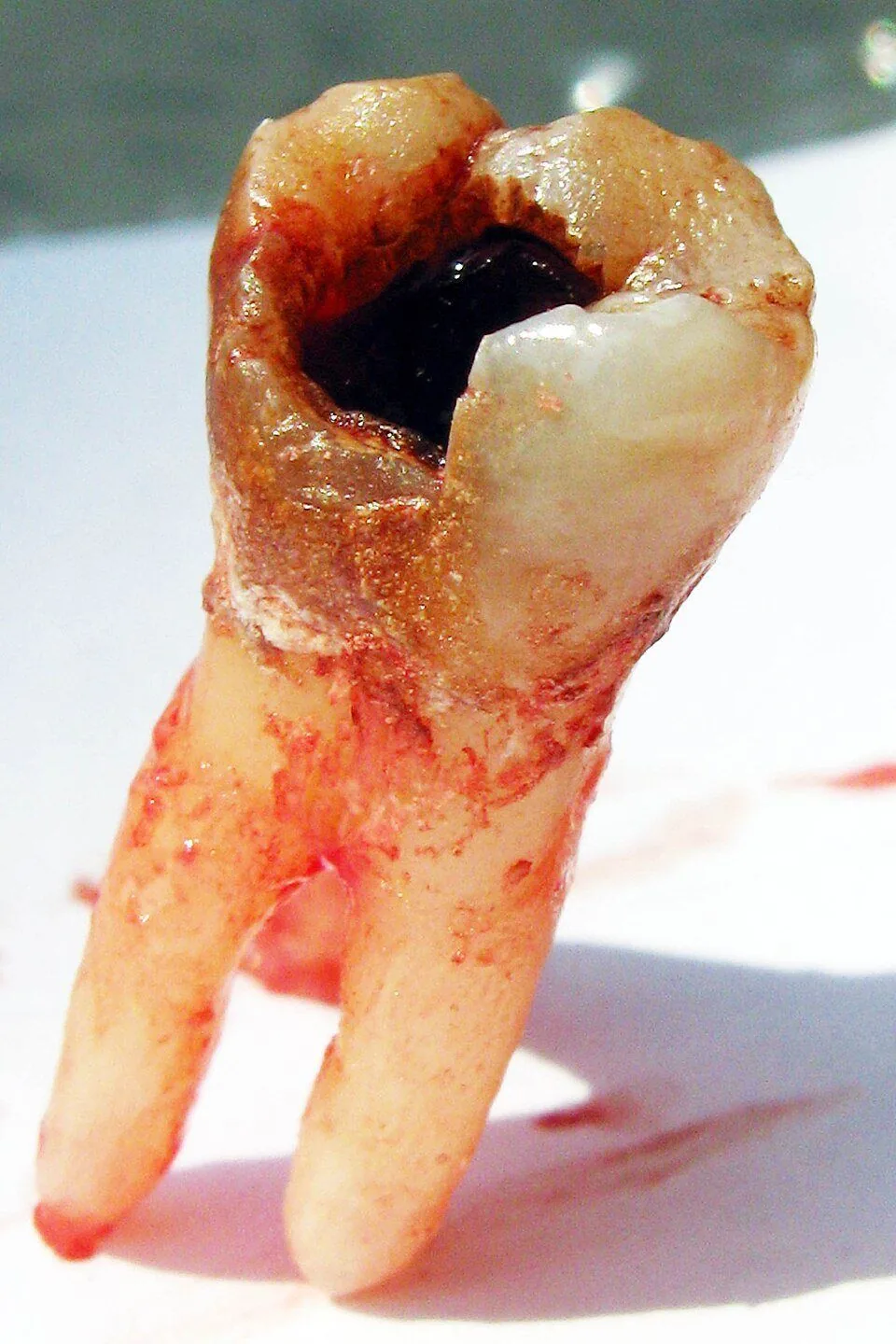
Simple extraction, step by step
In a simple extraction, you break the periodontal ligament and expand the socket with elevators, and then remove the tooth with forceps.
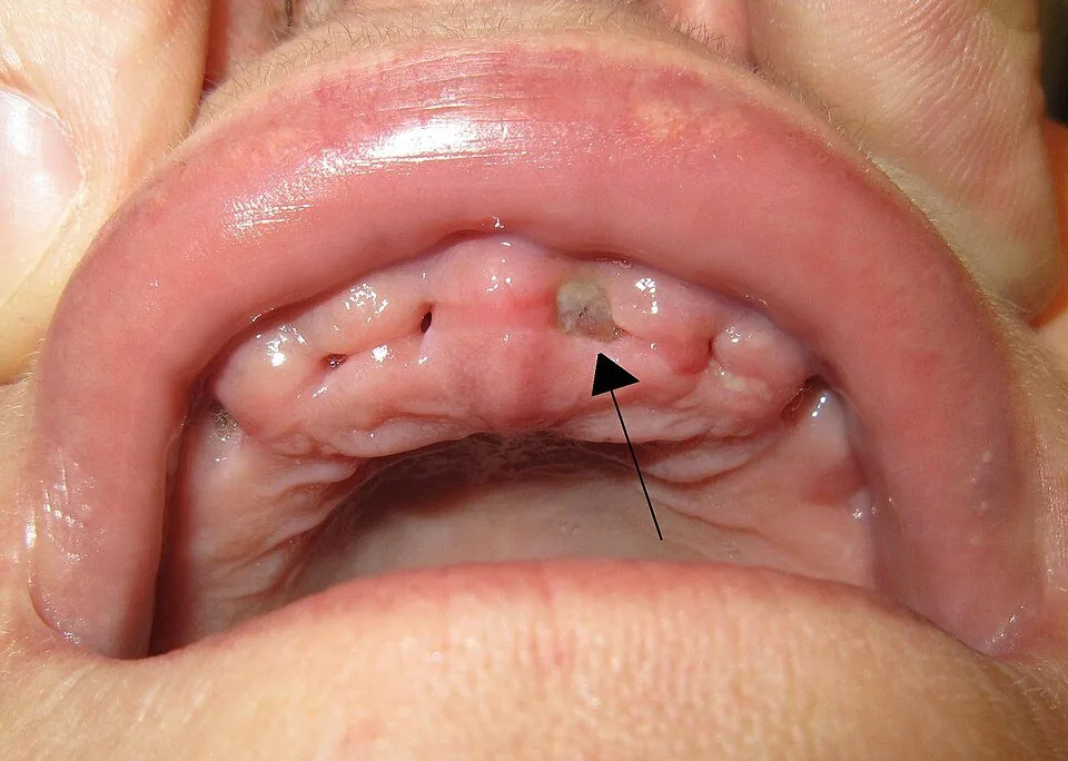
Healing after an extraction
After an extraction, the socket fills with blood clot → granulation tissue → immature bone → mature bone.
Impacted third molars: Winter and Pell & Gregory
A third molar (wisdom tooth) is impacted when it doesn't fully erupt because it runs into another tooth, the bone or the gums.
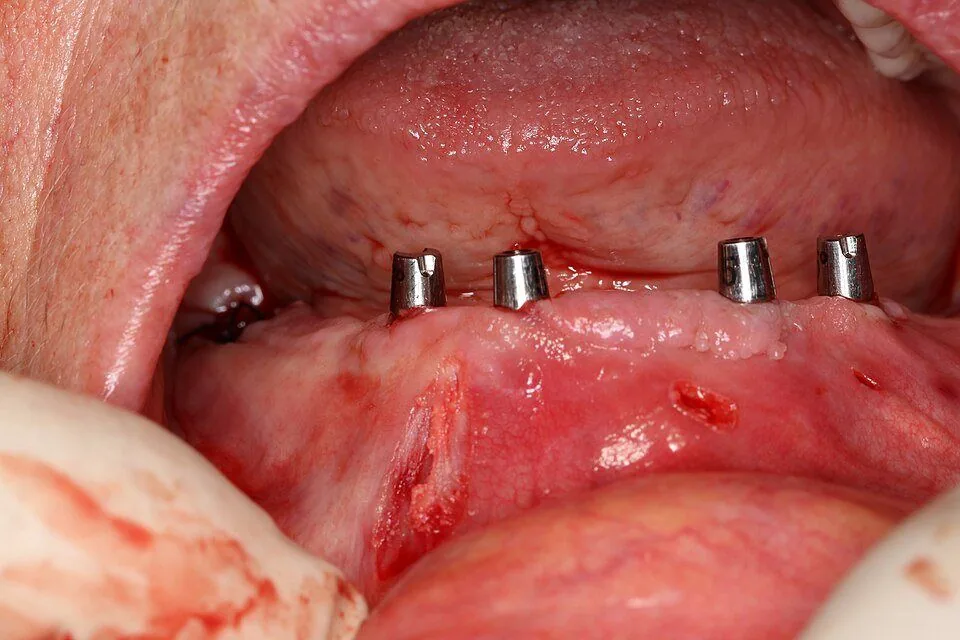
Osseointegration
Osseointegration is the direct, structural and functional connection between living bone and the surface of an implant that bears load (the force of chewing).
Play until it comes without thinking. Your best score is saved on your phone.
The games need JavaScript turned on in your browser.
Odonto Fácil on your phone
Install it like an app: it stays on your home screen, opens full screen and the notes work offline. Free.